
Pro-Dense Injectable Regenerative Graft is a bone graft substitute that is a purely synthetic composite of calcium sulfate and calcium phosphate. These two materials are biocompatible and have been used in orthopedics for over 100 years. The formulation in Pro-Dense Graft, however, is unique and has demonstrated excellent results in benign bone cysts and tumors.1-3
Pro-Dense Graft is currently the only bone graft substitute on the market to be indicated specifically for use in benign bone cysts and tumors in patients 6 years and up. The product allows for an injection technique in simple or unicameral bone cysts, or as a backfill in tumors/cysts in open techniques.
How is Pro-Dense Graft applied?
A generalized surgical technique is demonstrated below:
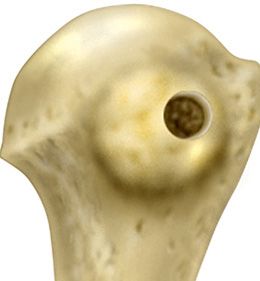
Step one
Localize the lesion under fluoroscopy. Create cortical access to facilitate curettage.
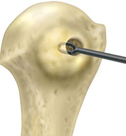
Step two
Using an image intensifier, excise the lesion, using a curette to debride defect margins. Excised tissue should be sent to pathology for routine histological examination and culture.
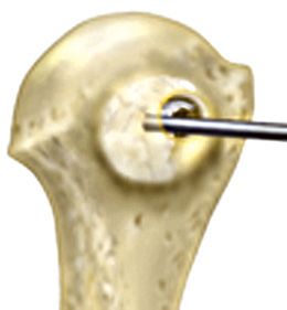
Step three
Having debrided the defect area, use the included delivery needle to inject Pro-Dense Graft into the defect.
What happens after application?
After implantation, the bone graft resorbs over time and allows the body to lay down dense new bone. This is different than other bone graft materials that take much longer (sometimes years) to resorb and be replaced by bone. In most cases, Pro-Dense Graft can be seen radiographically (i.e., on X-ray or CT) during postoperative visits. The surgeon will define postoperative activity level as the material resorbs and new bone heals and replaces the graft. The below case examples demonstrate how the material can be seen on X-ray and be replaced by dense new bone as the material is resorbed.
Case studies
Twelve-year-old male with a recurrent unicameral bone cyst of the right proximal humerus. Pro-Dense Graft was used to completely fill the lesion. No complications were reported. New bone formation was detected at 7 weeks, and at 7 months, a small amount of the residual graft was left. At 3 and 12 months, radiographs show dense new bone formation. Patient was fully active at 10 weeks and remains at full activity at present.
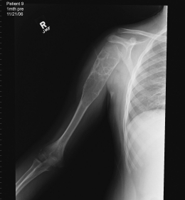
Preoperative
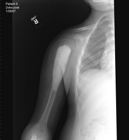
2 weeks postoperative
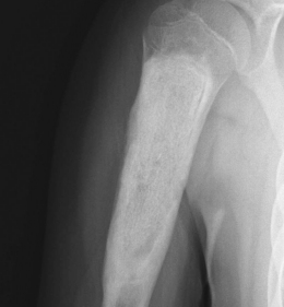
1 year postoperative
Images courtesy of: Robert Heck, MD - Campbell Clinic, Memphis, TN
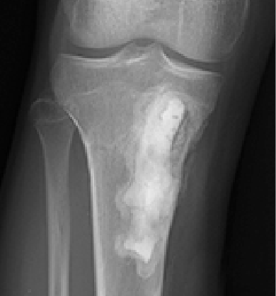
Sixteen-year-old male with a painful benign bone cyst of the right proximal tibia. The preoperative image shows the bone cyst measuring approximately 6cm x 3cm. It was debrided and grafted. The 1 month follow-up exhibits slight resorption of the graft and the patient was pain free with an independent activity level. The 3 month image shows further graft resorption and apparent dense bone regeneration. The 9-month radiograph demonstrates almost complete resorption of the graft and dense bone regeneration.
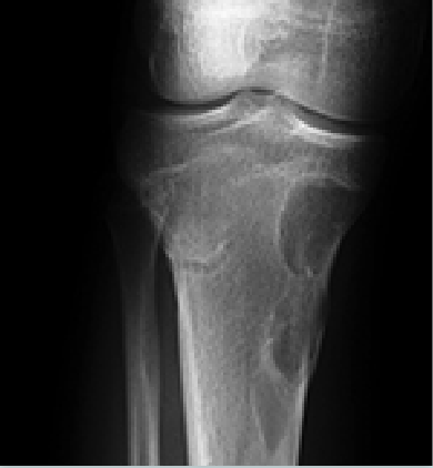
Preoperative
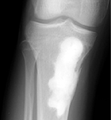
6 weeks postoperative
3 months postoperative
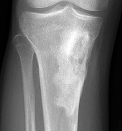
1 year postoperative
Images courtesy of: Mike Neel, MD - Ortho, Memphis, TN